|
Improving the optical resolution along the vertical direction for high speed 3D imaging of tissues |
|
Project 11 - Cihang (Sherry) Kong University of Bielefeld |
|
Objectives: (1) adaptation of high-speed GPU control and reconstruction routines for use with deep-tissue imaging by multi-focal SR-SIM (2) Extension of high-speed SR-SIM to 50 nm spatial resolution by non-linear structured illumination (laterally) and combination with fluctuation analysis (axially) (3) multi-color imaging of liver sinusoids in fixed rodent tissue by 2-photon fluorescence excitation based multifocal SIM (4) High-speed multifocal SR-SIM imaging of perfused liver slices |
|
Expected Results: (1) Imaging of LSEC dynamics in vitro (100 nm res., speed 60 reconstructed frames/s) (2) high resolution and high speed imaging of LSEC dynamics by nonlinear SIM (50 nm, 150 raw data fps) (3) Super-resolution imaging assay for small molecules, i.e. ethanol, paracetamol; nanoparticles; in perfused tissue established, initial screening |
|
Project Lead: Early Stage Researcher:
|
Cihang Kong was born in China in 1993. After finishing her high school education in 2011, she moved to Hong Kong SAR. Cihang has joined the DeLIVER-ITN project since December 2018. Currently she works in Bielefeld University under the supervision of Prof. Thomas Huser. Cihang got her bachelor’s degree in medical engineering from the University of Hong Kong in 2015. There she got trained in the microfluidics lab for her bachelor’s thesis ‘mimicking glaucoma on a microfluidic chip‘. After that, she began to pursue her PhD research in the department of electrical and electronic engineering. Her PhD thesis is about constructing ultrafast fiber laser sources for nonlinear optical microscopy. Cihang’s research aim in the DeLIVER-ITN project is to improve the optical resolution along the vertical direction for high speed 3D imaging of tissues. The first part is to construct a multi-focal SR-SIM platform which has 50-nm resolution in all directions. The second part is to adapt two-photon excitation for the multi-focal SIM system, which enables the deep SIM imaging of tissues such as perfused liver slices. |
P
|
Development of high-speed optical nanoscopy utilizing two-photon fluorescence excitation |
|
Project 10 - Sara Kheireddine LaVision Biotec GmbH |
|
Objectives: (1) Integration of confocal two-photon fluorescence microscopy with rescanning SIM and image inversion interferometry (2) utilize membrane deformable mirror optics to correct for wavefront distortions during deep tissue imaging (3) multi-color imaging of liver sinusoids in fixed rodent tissue by 2-photon SR-SIM utilizing adaptive optics wavefront corrections (4) high-speed 2-photon SR-SIM imaging of endothelial tubes, precision cut liver sections, and slices from histological preparations in perfusion chambers |
|
Expected Results: (1) Two-photon fluorescence microscopy with rescanning SIM and image inversion interferometry demonstrated (resolution 150 nm, speed: 8 frames/s) (2) imaging of 3D structure of LSECs in planar and 3D cell culture in microfluidic chambers (3) influence of small molecules (ethanol, paracetamol) and lipoprotein particles on fenestration stability imaged in perfused organ-on-chip models |
|
Project Lead: Early Stage Researcher:
|
Sara Kheireddine is a 28-year-old Canadian-Lebanese student who joined the DeLIVER ITN network in 2019 as an ESR in LaVision BioTec in Bielefeld, Germany. Born and raised in the United Arab Emirates, Sara then moved to Lebanon in 2008 to pursue her undergraduate studies at the American University of Beirut. After receiving her B.Eng. in Electrical and Computer Engineering, she moved to Germany in 2013 to do an MSc. in Biomedical Engineering at RWTH Aachen University. After finishing her Master thesis in McGill University in Canada, she got accepted into the Ph.D. program in 2016, and is currently a Ph.D. candidate in Bioengineering at McGill. Sara’s Ph.D. thesis is primarily concerned with visualizing biological agents in confined spaces, where she works on building and adapting various microscopy and imaging techniques to achieve high spatial resolution with a large field-of-view. At LaVision BioTec GmbH, Sara is working with light sheet fluorescence microscopy (LSFM) and two-photon microscopy to improve spatial resolution through enhanced multiview reconstruction, with the ultimate goal of achieving isotropic resolution. Moreover, structured illumination microscopy (SIM) will eventually be incorporated with two-photon microscopy for high resolution imaging of live samples. The end goal of these improvements is to realize the dynamic visualization of liver sinusoidal endothelial cells (LSECs) with high spatial resolution and a large field-of-view. In her spare time, Sara enjoys exploring new places, walking in nature, reading books, and watching movies and series. |
|
Assessing the histological diversity of liver sinusoidal endothelial cells by optical nanoscopy |
|
Project 9 - Lianne van Os Vrije Universiteit Brussel |
|
Objectives: (1) SR-SIM assessment of the morphology and diversity of LSECs in different liver diseases (e.g. cancer, autoimmune disease, alcoholic and non-alcoholic fatty liver disease) by isolating cells from human donor material and mouse models of liver injury (e.g. liver fibrosis, non-alcoholic fatty liver disease, cancer) (2) assess LSEC morphology and their interactions with other liver cells such as hepatic stellate cells and hepatocytes based on high-resolution imaging techniques (nanoscopy, EM) using immunohistochemistry of liver tissue and 3D hepatic spheroid cultures (3) heterogeneity of LSECs isolated from diseased liver tissue assessed by correlative molecular analysis techniques and SR-SIM with extended z-resolution |
|
Expected Results: (1) LSECS from human and/or mouse pathological liver specimens isolated, fixed, and sorted based on specific liver disease (2) morphology of isolated LSEC samples analyzed by SR-SIM (3) morphology and cellular identity confirmed by SR-SIM within the consortium and correlated to electron microscopy |
|
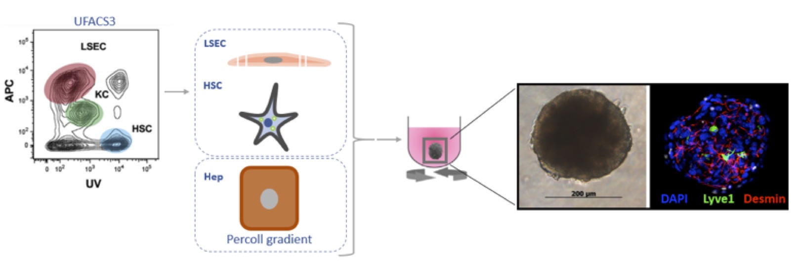
Globally, liver disease accounts for 2 million deaths per year of which cirrhosis, an advanced stage of fibrosis, causes 1.16 million deaths 1. Fibrosis is a response to chronic liver injury that can be initiated by several factors such as alcohol, non-alcohol fatty liver disease and viral hepatitis 2. LSECs play an important role in fibrosis 3-5and it is even suggested that LSECs could play a role in fibrosis initiation as the their specific phenotype changes (also known as capillarization) before fibrosis occurs in several liver diseases6, 7. LSECs line the sinusoidal wall and are characterized by the absence of a basal lamina and the presence of open pores also known as fenestrae that act as a dynamic filter8. During capillarization LSECs lose their fenestrations and obtain a basement membrane. Research aimed to elucidate mechanism involved in this capilarization of LSECs has focused on LSEC cultured in mono-layers whereby these cells capillarize due to the change to the artificial environment9-11. Co-culture of LSECs with other liver cell types or conditioned medium of other liver cells can improve LSEC function in culture 10, 12. However, to study LSECs in both healthy and diseased conditions, improvements need to be made for in vitroculture of LSECs. Therefore, we aim to develop a 3D spheroid culture with LSECs, hepatocytes and HSCs 13, 14to unravel the morphology and diversity of LSECs in both healthy as diseased states. In this case, the health status of LSECs can be determined by the expression of functional markers and fenestrae assessment by conventional Electron Microscopy (EM) and Super Resolution Microscopy (SRM) that will be developed by our consortium. Diseased states can be induced by for example Acetaminophen (for hepatocyte damage) or Monocrotaline (for LSEC damage) administration. In parallel, to get a complete overview on LSEC physiology in different liver etiologies, LSEC presence and morphology in different mouse models of liver disease will be investigated by immunohistochemistry and SRM of paraffine imbedded liver tissue.
Figure1: Schematic representation of spheroid formation procedure. HSCs and LSECs are isolated via the UFACS314 procedure and hepatocytes by a Percoll gradient. The freshly isolated cells are allowed to form spheroids in 96-well round bottom plate with a cell-repellent surface. After 1h plates are placed on an orbital shaker for the entire culture. Spheroid formation is already seen after 24h. Here a spheroid is shown at 5 days of culture and is stained for Lyve1(LSEC), Desmin (HSC) and DAPI (nucleus). References:
|
|
Project Lead: Early Stage Researcher:
|
During my bachelor Biomedical Sciences at the University of Utrecht (2012-2015) my interest started for research in disease modeling and liver research. With that motivation I started the master Biology of Disease at the University of Utrecht (2015-2017) where I focused on copper storage disease modelling in liver organoids at the Faculty of Veterinary Medicine at the lab of Prof. Bart Spee. For more insight into alternative sources for liver transplantation, I went to the Karolinska Institute to the lab of Prof. Stephen Strom were I worked on amnion epithelial stem cells as therapeutic cell source for the Alagille syndrome (rare genetic disorder that results in liver disease). After obtaining my Master’s degree in 2017, I started my PhD in 2018 at the LIVR cell biology research group of Prof. Leo van Grunsven on the role of liver sinusoidal endothelial cells in chronic liver diseases.
|
P
|
Assessing signal transduction processes in LSECs under inflammatory conditions by optical nanoscopy |
|
Project 8 - Eva Arias Bravo University of Bielefeld |
|
Objectives: (1) SR-SIM investigation of signal transduction under inflammatory conditions using a transgenic mouse reporter for NF-kB to assess stress of isolated LSECs (2) High-speed SR-SIM imaging to determine the effects of NF-kB inhibitors (Aspirin, glucorcorticoids), monitor all NF-kB DNA binding subunits (p50, p65, c-Rel, RelB and p52), and to follow inhibition of nuclear translocation of NF-kB by Cineol (3) utilize combined 2-photon, high speed SR-SIM and microfluidic perfusion of liver slices to assess NF-kB inflammation markers in liver tissue under environmental control and environmental stress, i.e. hypoxia |
|
Expected Results: (1) LSECs and liver slices isolated from reporter mouse, NF-kB activation monitored by SR-SIM (2) NF-kB inhibitor and activators in LSECs and liver slice cultures assessed (3) distribution and dynamics of NF-kB DNA binding subunits in primary cells assessed (4) 2-photon SR-SIM characterization of distribution of inflammation markers throughout perfused liver tissue completed |
|
Project Lead: Early Stage Researcher:
|
Eva Arias completed a Bachelor’s degree in Biology from the Complutense University of Madrid, Spain. Later, she completed a Master´s degree in Microbiology at the University of Alcalá in Madrid, Spain. During the Bachelor’ and Master’s thesis, she worked with Strongyloides stercoralis, a human pathogenic parasite causing the disease strongyloidiasis. Currently, Eva is the ESR number 8, and her project title is “Assessing Signal Transduction Processes in Liver Sinusoidal Endothelial Cells (LSECs) under Inflammatory Conditions by Optical Nanoscopy”. The main themes of her work are:
|
Page 2 of 4