ESR 1 (University of Bielefeld) - Jakub Pospisil
ESR 2 (University of Oxford) - Ana Rita Faria
ESR 3 (University of Tromsø) - Nikhil Jayakumar
ESR 4 (Elvesys) - Alessandra Dellaquila
ESR 5 (University of Tromsø) - Jennifer Cauzzo
ESR 6 (University of Gothenburg) - Zofia Korczak
ESR 7 (Cherry Biotech) - Matteo Boninsegna
ESR 8 (University of Bielefeld) - Eva Arias Bravo
ESR 9 (Vrije Universiteit Brussel) - Lianne van Os
ESR 10 (LaVision BioTec) - Sara Kheireddine
ESR 11 (University of Bielefeld) - Cihang Kong
ESR 12 (University of Birmingham) - Pantelitsa Papakyriacou
|
Super-resolution imaging of human LSECs in 3D culture and organ slices |
|
Project 14 - Karolina Szafranska University of Tromsø - The Arctic University of Norway |
|
Objectives: (1) Optimisation of endothelial tube formation in 3D cell culture (2) SR-SIM imaging of living LSECs forming endothelial tubes (3) optimization of staining and SR-SIM imaging of living endothelial tubes, correlation to perfused liver slices in perfusion chambers (4) SR-SIM and multifocal 2-photon SR-SIM imaging of endothelial cell interactions with small molecules and NPs in perfused sinusoids |
|
Expected Results: (1) Local supply of human LSECs established (2) imaging of the 3D structure of LSECs in tubes and 3D cell culture in vitro completed (3) high speed SR-SIM of human LSEC dynamics in 3D cell culture established (4) SR-SIM imaging of interactions of small molecules and nanoparticles with LSECs in perfused sinusoids performed |
|
Project Lead: Early Stage Researcher:
|
Karolina Szafranska is a 26 year old Polish research student who moved to the Arctic to follow her dream of studying liver sinusoidal endothelial cells (LSECs). Karolina has been a PhD student at the University of Tromsø (UiT) since February 2018, working as an Early Stage Researcher in the Vascular Biology Research Group to study the effects of chemical compounds on LSECs. Karolina has Bachelor and Master degrees in Biophysics from the Jagiellonian University in Poland, where she focused on the application of atomic force microscopy to study the dynamics of the liver sieve on fenestrated liver endothelial cells. These results are published and presented at the 19th Liver Sinusoid Meeting in Galway, where Karolina has met her new scientific family from Tromsø. Since then, she is using her previous experience in combination with the long tradition of LSEC study and novel nanoscopy techniques at UiT. In her free time, Karolina enjoys life in the Arctic, skiing, cycling, removing snow from her driveway and developing Stockholm syndrome with the one and only (local) radio station. Karolina’s project as Early Stage Researcher in the DeLiver ITN has two main topics. The first part focuses on the development of co-culture of liver cells to create small, artificial livers in in vitro conditions. The second part is about studying effects of various drugs, substances or nanoparticles on liver endothelial cells. Combined knowledge from both parts of the project will hopefully lead to the development of a system for in vitro drug testing prior to human/animal trials. |
|
Optical nanoscopy of the pharmacological clearance function of LSECs |
|
Project 13 - Larissa Kruse University of Tromsø - The Arctic University of Norway |
|
Objectives: (1) Generation of monoclonal antibodies to stabilin-2, the major workhorse LSEC scavenger receptor (2) Use of these antibodies to map the fate of stabilin-2 and its ligands in LSEC during scavenging by SR-SIM (3) Use of antibodies to determine changes in the expression (number and location) of LSEC stabilin-2 in response to small molecule challenge in vitro and in vivo in rodents, and in precision cut liver slice specimens from animal models of liver disease and of diseased and normal human liver specimens by SR-SIM |
|
Expected Results: (1) Highly specific antibody reagents that recognise stabilin-2 generated (2) dissemination of antibodies to beneficiaries and partners (3) nanoscopic imaging of stabilin-2 dynamics in vitro demonstrated (resolution 100 nm, speed 20 frames/s (3D) (4) changes in stabilin-2 expression determined in response to small-molecule interactions (5) imaging of stabilin-2 within LSEC precision cut liver slices by SR-SIM nanoscopy achieved |
|
The project is about the liver sinusoidal endothelial cells (LSECs) and their scavenger function. The liver consists of different cells, which are the parenchymal and non-parenchymal cells. The liver cells that most people know about are the hepatocytes. Larissa is working with one family of the non-parenchymal cells, namely the liver sinusoidal endothelial cells or LSECs. LSECs are cells that line the sinusoids, which are the capillaries of the liver. The endothelial cells line all vessels that direct the blood running in your body. In comparison to other endothelial cells, LSECs are special because they have holes called "fenestrations". These «liver holes» are found in groups called sieve plates and they are dynamic structures. That means they can be bigger and smaller and they can also disappear completely. The fenestrations become smaller and fewer with age, liver diseases such as fibrosis and through substance abuse such as excessive alcohol consumption. LSECs filter substances and fluids out of the blood stream. This process is called “scavenging”. Some substances might be able to change the “holiness” of the LSECs and influence their scavenging process/behaviourand we want to know which substances are influencing the liver holes how. Larissa is trying to figure out, what makes liver holes happy. At the moment Larissa is working on the influence of branched chain amino acids (BCAAs) on LSECs. BCAAs are the amino acids leucine, isoleucine and valine. They belong to the essential amino acids and therefore need to be taken up by the human body, for example, with food. BCAAs are known to be taken by athletes and body-builders as they are said to help with muscle built up and they are supposed to give you more energy. Larissa is interested in how the BCAAs influence the LSEC fenestrations and she would like to see what is happening. Therefore she is working with endocytosis experiments but also with super resolution microscopy to make the holes visible for the naked eye. Larissa is using scanning electron microscopy (SEM), structured illumination microscopy (SIM) and direct stochastic optical reconstruction microscopy (dSTORM). The goal is to find targets and develop antibodies for certain receptors to make the functionality of the LSEC visible and thus be able to find out how the different substances, for example BCAAs influence the LSEC fenestrations. |
|
Project Lead: Early Stage Researcher:
|
Chocolate and book-loving German village nerd-kid travelling to find her research home in Arctic vascular biology This is Larissa Kruse. She is 28 years old and originally from Germany. Larissa started studying Forensic Sciences in Bonn, Germany in 2010. Since then, she has moved from there to Scotland and England to study some more Forensic Science and Ballistics. In 2015, she came back to the north of Germany, Bremerhaven, where she did her M.Sc. in Biotechnology. During her thesis, which she wrote in Stockholm, Sweden, Larissa did research in medical biology and proteomics and this is her professional field of interest now. Larissa likes nature, especially animals and water and loves reading books and watching movies. In September 2018 Larissa moved to Tromsø, Norway, which is way above the Arctic Circle and therefore is not displayed on German weather maps. Larissa is a PhD student in the Vascular Biology Research Group and part of the DeLIVER project. |
|
Development of perfused human liver slice model for optical nanoscopy |
|
Project 12 - Pantelitsa Papakyriacou University of Birmingham |
|
Objectives: (1) To develop a human precision cut liver slice model for imaging live human LSECs in normal and cirrhotic tissue specimens (2) To adapt slicing system to permit intravascular perfusion of scavenger ligands and monoclonal antibodies, integration with organ-on-chip perfusion chambers (3) To optimise culture and fixation protocols to permit temporal assessment of LSEC by high-speed SR-SIM imaging, 2-photon SIM imaging of perfused liver slices |
|
Expected Results: (1) methodology to permit dynamic visualization of cellular fenestrations in human LSEC and assess alterations by capillarisation and cirrhosis developed (2) comprehensive assessment of the expression and subcellular localisation of scavenger receptors such as L-SIGN, Stabilin -1 and -2, CD36, Mannose receptor and FcR’s on human LSEC in situ (3) detailed temporal analysis of the endocytic capabilities of human LSECs with respect to uptake of small molecules and lipoproteins (4) optimised methodology for generation and culture of human precision cut liver slices for imaging modalities developed |
|
Project Lead: Early Stage Researcher:
|
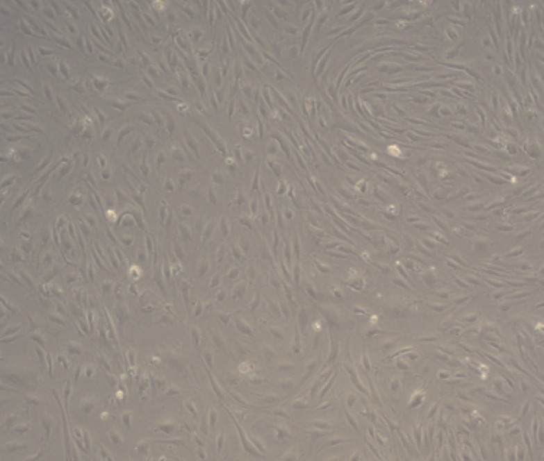
Pantelitsa completed her BSc in Biomedical Sciences at Coventry University, UK. Following this she completed two master degrees, one in Molecular Genetics and Diagnostics and secondly Biomedical Research. Her DeLIVER project will harness the human liver tissue samples and cell isolation protocols available in Birmingham (Figure 1) to characterise the structure and function of fenestrations. These contribute to the molecular sieve function of human liver sinusoidal endothelium, and will be studied in the context of states of disease as well as ageing. These sinusoidal cell fenestrations are dynamic structures, and along with the expression of unusual scavenger receptors on the endothelium, facilitate the scavenging functions of these cells. This is important because maintenance of proper function of the endothelial nanopores is crucial to homeostasis of lipid metabolism and for immune surveillance. In chronic liver disease pseudocapillarisation occurs whereby liver endothelium is defenestrated and a more mature basement membrane rich in collagen develops. This process also takes place during ageing and this has serious implications as defenestration can compromise the efficient absorption of nutrients, exchange of materials synthesized by the liver and clearance of drug treatments. Pantelitsa’s project will use human liver sinusoidal endothelial cells isolated from normal and diseased liver specimens to compare the organisation and function of fenestrations and understand how the disease process regulates their expression. We will also study cells in situ in intact tissue wedges and will use both systems to assess the effect of agents such as alcohol and specific growth factors such as VEGF on fenestration dynamics. Since the fenestrations can only be visualised at the nanoscale, Super Resolution Imaging as well as Electron Microscopy will be implemented on cells derived from explanted liver tissue specimens. We hope to optimise our imaging protocols to obtain a spatially resolved image of the fenestrations in different samples.
Figure 1: Representative phase contrast image of human hepatic sinusoidal endothelial cells isolated from an Alcoholic Liver Disease sample. |
Page 1 of 4


