
|
Development of a waveguide- and microfluidics-based integrated optical nanoscopy platform |
|
Project 2 - Nikhil Jayakumar University of Tromsø - The Arctic University of Norway |
|
Objectives: (1) Development of an integrated optics platform for real-time nanoscopic study of LSEC dynamics based on waveguide-SR-SIM (2) Increase field-of-view for single molecule localization microscopy to 200 µm x 100 µm based on waveguide excitation, implement nonlinear SIM approaches (3) demonstrate waveguide-integrated nanoscopy with microfluidic delivery for the rapid imaging of small molecule-LSEC interactions |
|
Expected Results: (1) Waveguide nanoscopy operational and imaging of fixed cells completed (90 nm, 20 frames/s) (2) field of view of waveguide nanoscopy increased to 200 µm x 100 µm (3) imaging of interactions of small molecules and nanoparticles with LSECs in vitro demonstrated |
|
Project Lead: Early Stage Researcher:
|
Nikhil Jayakumar was born in Kerala, India. He completed his Bachelors degree in Electrical and Electronics engineering from the University of Kerala and then worked under different verticals before moving to Jena, Germany in 2015. In Jena he graduated with a Masters degree in Photonics in 2017 and continued working as a scientific assistant before moving to UiT, Tromso for his Ph.D. He started his work in November 2018 under the supervision of Assoc. Prof. Dr. Balpreet Singh Ahluwalia and the scope of his work includes super-resolution imaging using waveguides and quantitative phase microscopy.
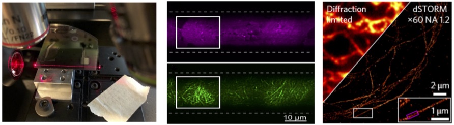
Nikhil Jayakumar's project will involve chip-based optical nanoscopy and quantitative phase microscopy. Chip-based optical nanoscopy: Our group have recently developed photonic chip based optical microscopy and nanoscopy methodologies [1, 2]. The use of a waveguide chip has enabled miniaturization of the optical setup by decoupling the excitation and collection paths (see Fig. 1a, below). The sample to be imaged is placed on top of the waveguide core area and the evanescent field generated due to total internal reflection at the core-sample interface illuminates. Due to evanescent field excitation only, a thin portion of the sample (a few hundred nm) is illuminated providing a very high signal-to-noise ratio TIRF images as shown in Fig. 1b. By using waveguide made of high refractive index contrast and by using thin waveguides the intensity of the evanescent field can be enhanced enabling blinking of fluorophores and the demonstration of on-chip optical nanoscopy using dSTORM principles as shown in Fig. 1(c) [2].The field of view (FOV) of the chip-based optical nanoscopy can be made large and we have recently reported optical resolution of 70-75 nm over FOV of 500 x 500 μm2 [3]. Nikhil Jayakumar's project will further work upon this novel idea of high throughput chip-based nanoscopy. Currently dSTORM imaging is limited by temporal resolution (takes approximately 30 minutes), as a large number of images (more than 20000 images) is required for a faithful reconstruction of the image. We intend to bring down this temporal resolution to a few seconds by employing intensity-fluctuation based algorithms such as MUSICAL, SOFI, ESI etc. which will facilitate high throughput imaging of liver sinusoidal endothelial cells. Another task is to build a portable photonic- chip based optical nanoscope and distribute within DeLIVER network.
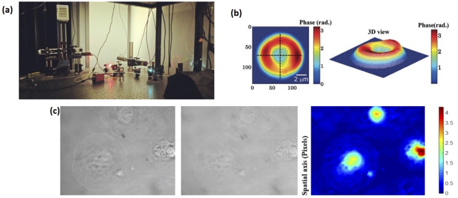
Figure 1: (a) Silicon nitride waveguide on a chip guiding light at 660 nm (b) TIRF image of stained MCC13 cells illuminated using a 25 μm wide silicon nitride waveguide (figure taken from Ref. 2) (c) Immuno-stained tubulin in LSEC under diffraction-limited imaging and d-STORM on waveguide based imaging (figure taken from Ref. 1) Quantitative phase microscopy: Biologists prefer to study cells living in their natural environment. However, most optical microscopic techniques rely on labeling the sample which is then no longer a natural state of the cell. And that is where quantitative phase microscopy (QPM) becomes useful. QPM provides a non-invasive non-contact imaging technique which can provide quantitative information like refractive index, thickness of the cell etc. It is known that when light passes through a sample that is weakly absorbing, which is the case for most cells, its phase changes. This change in phase contains information about the refractive index and thickness of the cell. The beauty of QPM is that to decode the information about the cell structure, the light beam which contains information about the cell needs to interfere with a reference beam, from which the relative change in phase can be deduced thereby ensuring label-free imaging. As part of this Ph.D. work, considerable emphasis will be laid on developing new techniques using the principles of QPM and improve the transversal and longitudinal resolution of QPM. By increasing the spatial resolution and temporal sensitivity of the novel quantitative phase microscopy, we aim to perform label-free quantitative imaging of the nanoscale fenestration present on the cell membrane of the LSECs. To enhance the spatial resolution of QPM, we will explore novel oblique angle illumination and Fourier ptychography imaging methodologies. And for enhancing the temporal sensitivity we will work with a common path approach. Fig. 2a shows the newly developed QPM at UiT and the results obtained from red blood cells (Fig. 2b) and initial results obtained from LSECs (Fig. 2c).
Figure 2: (a) Quantitative Phase Microscopy setup at UiT, a Linnik interferometer is used for the generation of interference fringes (b) 2D and 3D images of Red Blood Cells phase informatin obtained using QPM (figure taken from Ref. 4) (c) Bright field image of LSEC (A5), interferometric image of the same location and the reconstructed phase map of the similar cell. Bar represents the phase map “𝜙”in radian References: [1] - Diekmann et et.al. Chip-based wide field-of-view nanoscopy, Nat. Photonics 2017 doi: 10.1038/nphoton.2017.55 [2] - Tinguely, et.al. Silicon nitride waveguide platform for fluorescence microscopy of living cells, Opt. Express 2017; Volume 25 (22). doi:10.1364/OE.25.027678 [3] - Helle, et.al., Nanoscopy-on-a-chip:super-resolution imaging on the millimeter scale, Opt. Express 2019; Volume 27 (5). doi.org/10.1364/OE.27.006700 [4] - Ahmad, et.al., Quantitative phase microscopy of red blood cells during planar trapping and propulsion, Lab on a Chip 2018; Volume 18 (19). doi: 10.1039/c8lc00356d |
|
Adaptive optics 3D-AO-SIM for fast 3D super-resolution SIM imaging of fixed and live tissues |
|
Project 2 - Ana Rita Faria University of Oxford |
|
Objectives: (1) Develop a low cost, portable SR-SIM solution (2) Implement adaptive optics (developed at the Booth Lab, Oxford) to an upright SIM setup (Deep-SIM) developed at the Micron Oxford Advanced Bioimaging Unit (3) Apply 3D-AO-SIM imaging to precision cut liver slices (4) Image cells within tissue and their interaction with small molecules, nanoparticles in perfused, precision cut liver slices by 3D AO-SIM |
|
Expected Results: (1) Deep-SIM setup with adaptive optics and fast SLM pattern generation operational to enable aberration-free 3D-SIM imaging of fixed cells demonstrated (<150nm xy <400 nm z-resolution) (2) high resolution and high speed SR-SIM imaging of 3D structure of LSECs in 3D cell culture in vitro demonstrated (1 3D fps) (3) interactions of small molecules and nanoparticles with LSECs in perfused sinusoids demonstrated |
|
Project Lead: Early Stage Researcher:
|
Ana Rita Faria is a Portuguese DPhil student in the Biochemistry Department of the University of Oxford under the supervision of Dr. Lothar Schermelleh. She started her project in October 2018 as an Early Stage Researcher within the DeLIVER network.
Before moving to the UK, Rita was studying Biomedical Engineering at the University of Minho (Portugal) as part of a Master Internship Programme of the International Iberian Nanotechnology Laboratory (INL). In her Masters project, she was researching anticancer drug delivery systems using advanced fluorescence spectroscopy and imaging techniques.
Rita’s project within the DeLIVER network is focused on establishing the application of adaptive optics implemented on a bespoke upright structured illumination microscope (Deep-SIM)developed at the Micron Oxford Advanced Bioimaging Unit. This setup will allow aberration-free 3D super-resolution imaging in extended depth of up to 50 µm, thus enabling the study of 3D cell culture and tissues. The main aim is to image liver sinusoidal endothelial cells (LSECs), their fenestrae dynamics and interaction with small molecules and nanoparticles.
When she’s not in the lab, you will find her enjoying the (more often than expected) sunny days that Oxford has to offer, going for weekend trips with lots of hiking, camping and climbing.
|
|
Development of high-speed structured illumination microscopy with instant, GPU-based image reconstruction |
|
Project 1 - Jakub Pospíšil University of Bielefeld, Germany |
|
Objectives: (1) Utilize spatial light modulators and/or digital mirror devices to provide SIM pattern formation at rates of up to 60 Hz (2) extend open source SR-SIM image reconstruction software "fairSIM" to full 3D image reconstruction, port to GPUs for instant reconstruction, and implement on a low-cost platform (3) characterize structure and dynamics of fenestrated cells isolated mice, rats and humans (4) implement nonlinear SIM image reconstruction in fairSIM |
|
Expected Results: (1) fairSIM for 3D image reconstruction released as open source plug-in for Fiji (2) fairSIM ported to GPUs, running at 60 reconstructed frames/s (3) fairSIM controlling low-cost SIM system available as blueprint (4) nonlinear SIM reconstruction implemented in fairSIM |
|
Project Lead: Early Stage Researcher:
|
Jakub Pospíšil was born in Jinřichův Hradec, Czech Republic in 1990. In 2015, he received his master diploma from Communications, Multimedia and Electronics program at Faculty of Electrical Engineering (FEE), Czech Technical University (CTU) in Prague. Diploma Thesis: “Design of algorithm for segmentation of speech utterances in patients with Huntington's disease”. Jakub's main area of interest is super-resolution structured illumination fluorescence microscopy (SR-SIM) image processing and analysis for applications in cell biology. Jakub contributed to development of second generation of “SIMToolbox” – an open-source, modular set of functions for MATLAB designed for processing data acquired by structured illumination microscopy. Currently, he is particularly focused on the development of related advanced digital image processing algorithms. Jakub works on image quality assessment in structured illumination microscopy, especially on the analysis of reconstruction artifacts and assessing resolution in super-resolution reconstructed images. His main research objective in DeLIVER project is the development of high-speed structured illumination microscopy with instant, GPU-based image reconstruction. To this end, he will utilize spatial light modulators, digital mirror devices, and/or compact fiber-optic interferometers to generate interference patterns at rates of 60 Hz or more (WP1, Task 1.1). These patterns are used as excitation patterns for SR-SIM. He will also extend the existing open source SR-SIM image reconstruction software “fairSIM” to full 3D image reconstruction by porting the reconstruction algorithms to parallel-processing GPUs for instant image reconstruction. He will also attempt to implement a novel, compact SIM generation system on a low-cost microscope platform, which could be utilized for measurements during his secondments at partner sites (WP1, Tasks 1.2, 1.3). Once the development of these SR-SIM platforms is completed, he will characterize the structure and dynamics of fenestrated cells from isolated mice, rats and humans (WP1, Task 1.4). To obtain even higher spatial resolution below 100 nm, he will also attempt to implement nonlinear SIM image reconstruction algorithms in fairSIM (WP2, Task 2.1). |

Page 4 of 4