|
Development of perfused human liver slice model for optical nanoscopy |
|
Project 12 - Pantelitsa Papakyriacou University of Birmingham |
|
Objectives: (1) To develop a human precision cut liver slice model for imaging live human LSECs in normal and cirrhotic tissue specimens (2) To adapt slicing system to permit intravascular perfusion of scavenger ligands and monoclonal antibodies, integration with organ-on-chip perfusion chambers (3) To optimise culture and fixation protocols to permit temporal assessment of LSEC by high-speed SR-SIM imaging, 2-photon SIM imaging of perfused liver slices |
|
Expected Results: (1) methodology to permit dynamic visualization of cellular fenestrations in human LSEC and assess alterations by capillarisation and cirrhosis developed (2) comprehensive assessment of the expression and subcellular localisation of scavenger receptors such as L-SIGN, Stabilin -1 and -2, CD36, Mannose receptor and FcR’s on human LSEC in situ (3) detailed temporal analysis of the endocytic capabilities of human LSECs with respect to uptake of small molecules and lipoproteins (4) optimised methodology for generation and culture of human precision cut liver slices for imaging modalities developed |
|
Project Lead: Early Stage Researcher:
|
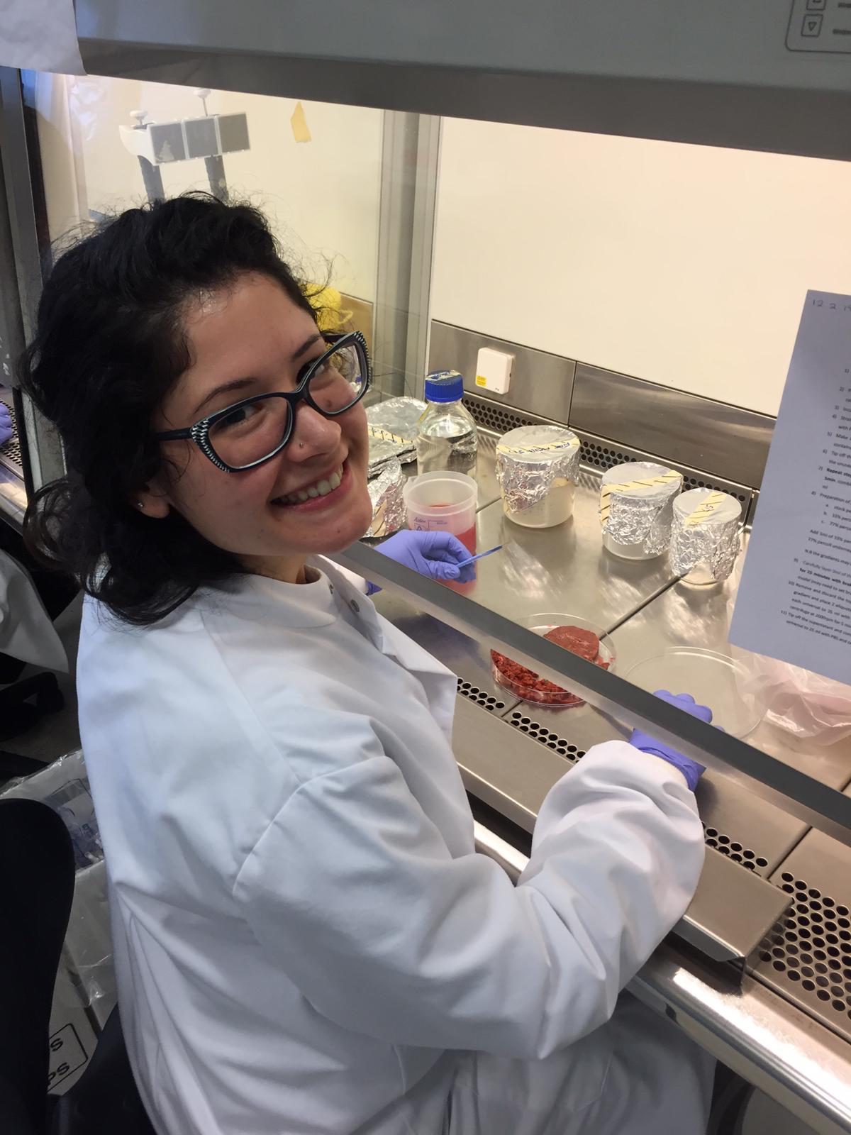
Pantelitsa completed her BSc in Biomedical Sciences at Coventry University, UK. Following this she completed two master degrees, one in Molecular Genetics and Diagnostics and secondly Biomedical Research. Her DeLIVER project will harness the human liver tissue samples and cell isolation protocols available in Birmingham (Figure 1) to characterise the structure and function of fenestrations. These contribute to the molecular sieve function of human liver sinusoidal endothelium, and will be studied in the context of states of disease as well as ageing. These sinusoidal cell fenestrations are dynamic structures, and along with the expression of unusual scavenger receptors on the endothelium, facilitate the scavenging functions of these cells. This is important because maintenance of proper function of the endothelial nanopores is crucial to homeostasis of lipid metabolism and for immune surveillance. In chronic liver disease pseudocapillarisation occurs whereby liver endothelium is defenestrated and a more mature basement membrane rich in collagen develops. This process also takes place during ageing and this has serious implications as defenestration can compromise the efficient absorption of nutrients, exchange of materials synthesized by the liver and clearance of drug treatments. Pantelitsa’s project will use human liver sinusoidal endothelial cells isolated from normal and diseased liver specimens to compare the organisation and function of fenestrations and understand how the disease process regulates their expression. We will also study cells in situ in intact tissue wedges and will use both systems to assess the effect of agents such as alcohol and specific growth factors such as VEGF on fenestration dynamics. Since the fenestrations can only be visualised at the nanoscale, Super Resolution Imaging as well as Electron Microscopy will be implemented on cells derived from explanted liver tissue specimens. We hope to optimise our imaging protocols to obtain a spatially resolved image of the fenestrations in different samples.
Figure 1: Representative phase contrast image of human hepatic sinusoidal endothelial cells isolated from an Alcoholic Liver Disease sample. |